- About the spectrometer
- Applications
- Other
- Accessory
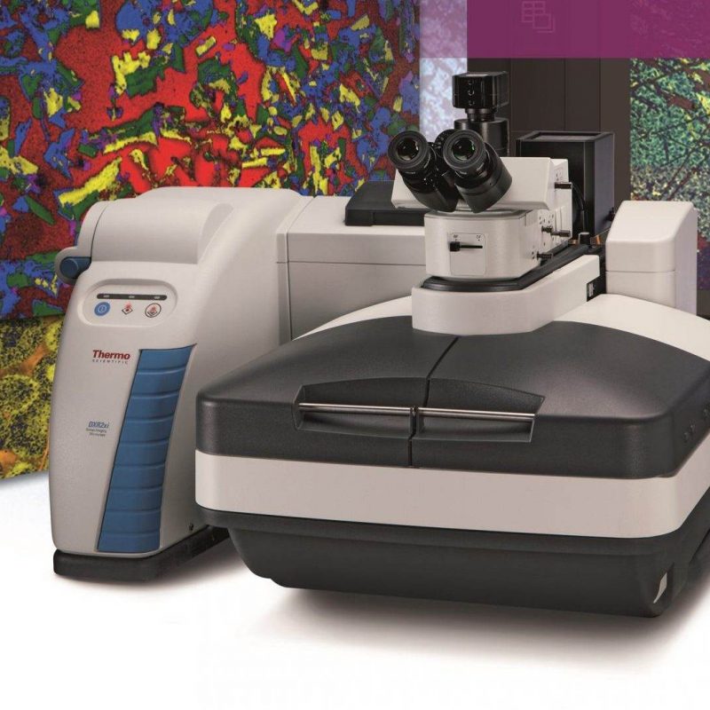
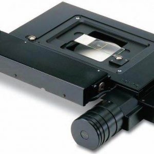
The new Thermo Scientific DXR3xiRaman microspectrometer takes Raman microscopy to a whole new level. Completely new solution for synchronizing the movement of a piezoelectrically controlled microscope stage with an EMCCD detector is unigue. All system components (lasers, gratings, filters) are user-replaceable and automatically detectable, the control program also allows the optimization of all system parameters in real time. The combination of these factors leads to an intensive simplification of the analysis of all types of materials by Raman micro-spectroscopy.
- Autoexposure and autofocus – like digital cameras. No more searching for optimal parameters and measuring with the method: “trial and error”!
- 3D visualization software, Particle Analysis function for microplastic analysis, etc.
- Spatial resolution: 540 nm in the X and Y axes, depth resolution approx. 1.7 micrometers (Z axis).
- More than five different excitation lasers for optimal acquisition of a spectrum of difficult samples.
- Laser power regulator for constant laser power delivered on the sample.
- Confocal design, excellent visual quality. Reflective and transmission exposure of the sample, fluorescent illumination.
- Patented quick automatic adjustment system for maximum performance.
- Fast, automatic multipoint calibration for confidence in sample identification: DynaCal automatic X-axis calibration.
- Compatibility with many high-quality Olympus microscopic parts.
- Class 1 laser safety – no workplace modifications required.
- Complete package of DQ/IQ/OQ/PQ validation protocols + CFR 21 part 11 compliance.
- Automatic polarization available.
- Possibility of combination of Raman and electron microscopy Raman-SEM
The novelty is especially the unrivaled speed of data acquisition, in a few tens of minutes it is possible to obtain millions of Raman spectra and with a resolution of 0.5 micrometers to map, for example,the whole pharmaceutical tablet! The DXR3 and DXR3xi Raman microscopes can also be quickly combined with AFM microscopes from various manufacturers to obtain information on the structural and topographic properties of the material surface, in nanometer units. Of course, there is also the possibility of TERS, SPM, etc. The combination of measuring techniques of Raman microscopy and AFM is a logical solution, e.g. for material engineering and other fields examining the surfaces of various materials.
Another option is a combination of Raman and electron microscopy: Raman-SEM correlative microscopy. Do not hesitate to contact us for more information.
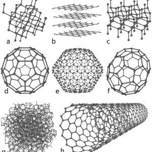
- Mapping of vapor-deposited graphene using a DXR3xi microscope
- Protective properties of graphene layers
- Mapping of geological materials using Raman microscopy
- Chemical mapping of tablet surfaces and study of component distribution
- Study of polymorphs of drug substances in low concentrations
- In situ analysis of Li-on batteries using Raman microscopy
- Ex situ analysis of Li-on batteries using Raman microscopy
