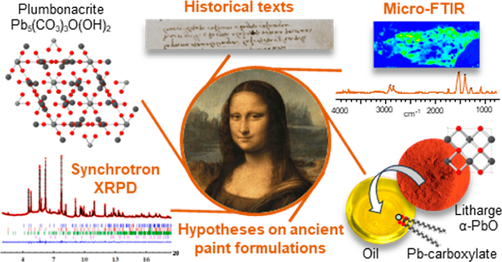
Leonardo da Vinci is undoubtedly one of the greatest geniuses of all time. Although more than 500 years have passed since his death, there is still much to discover in his works. One of his most famous creations is the legendary Mona Lisa. Even though it might seem that it is explored down to the last layer, it is not so, also for the reason that a number of analytical methods belong to invasive or destructive techniques.

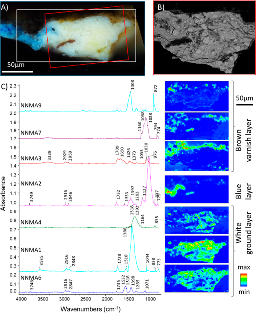
A team of European scientists managed to obtain an exceptional sample from the bottom layer of the image. This was analyzed using synchrotron X-ray diffraction and FT-IR microscopy, which is non-destructive and often it is not even necessary to take a sample from the studied object (also thanks to the large working distance and load capacity of microscope stages, which modern FT-IR microscopes offer). It was found that it contains a unique blend of high lead saponified oil and cerussite (PbCO3)-depleted lead white pigment. The most remarkable feature of the sample is the presence of plumbonacrite (Pb5(CO3)3O(OH)2), a rare compound that is only stable in an alkaline environment.

Leonardo probably prepared a thick paint suitable for covering the wooden panel of the Mona Lisa by treating the oil with a high content of lead oxide (PbO). However, his manuscripts draw ambiguous conclusions regarding the use of PbO. Analysis of fragments from The Last Supper confirms that not only PbO was part of Leonardo’s palette, but also litharge (α-PbO), massicote (β-PbO), plumbonacrite and shannonite (Pb2OCO3), the latter phase being detected for the first time in a historical image.
It turns out that the FT-IR microscope is an irreplaceable companion for every adventurer who wants to look under the hands of the old masters.
