+420 602 325 829 / info@nicoletcz.cz

Nano-imaging of nucleocyte nucleus

The neaSCOPE microscopesare designed for imaging and spectroscopy and for non-invasive analysis of the organization and morphology of biological samples with a spatial resolution of 10 nm.

DNA distribution in white blood cells

Nano-imaging of nucleocyte nuclei and analysis of measured spectra

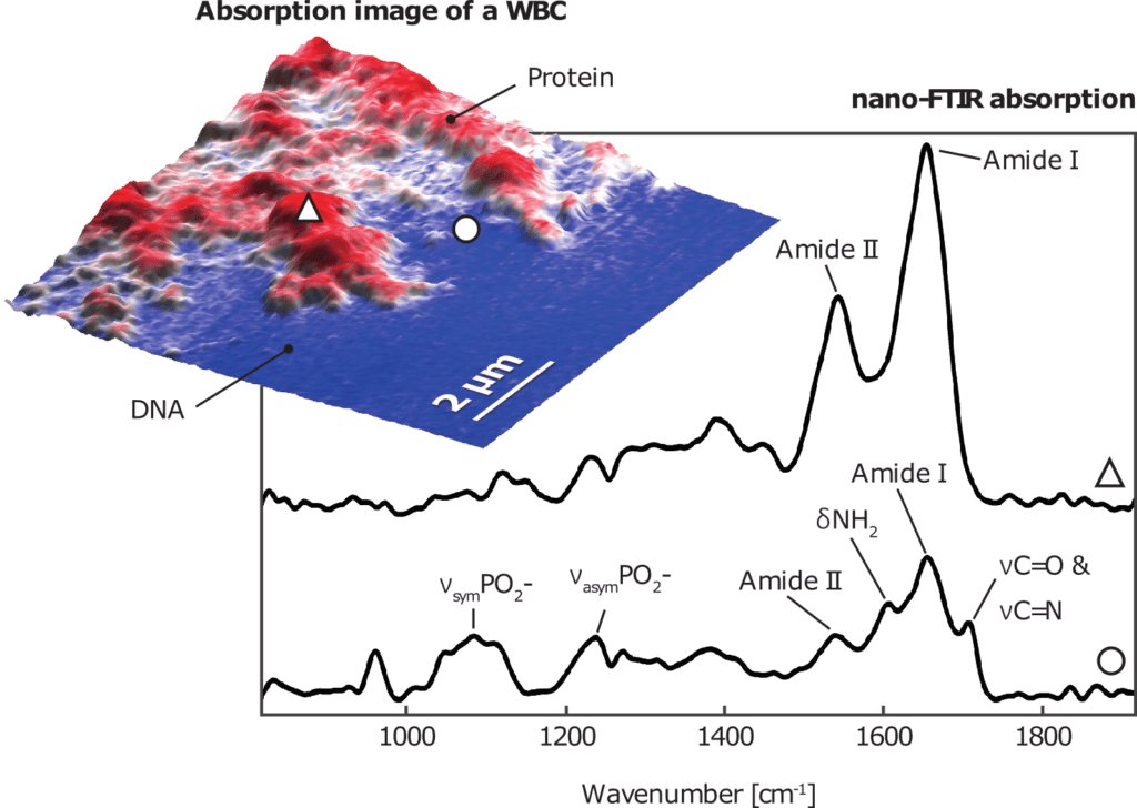
The IR image obtained with the neaSCOPE microscope shows a map of protein and DNA rich areas in white blood cells (WBC) overlaid by topography. nano-FTIR spectra in the thinner region (blue) show characteristic bands for single- and double-stranded DNA, indicating that this region is a nuclear pore. The thicker regions show no signs of nucleic acid bands, indicating protein-rich interphase chromosome regions, which are often associated with the development of diseases such as Hodgkin's lymphoma.

Learn more about this and other applications here.

IR-neaSCOPE is the basic model for infrared imaging and nano-spectroscopy. It provides maximum performance without damaging the sample. This is a cost-effective solution for samples with a high coefficient of thermal expansion.
IR-neaSCOPE+fs is designed for pump-probe spectroscopy with 10fs temporal and 10nm spatial resolution: it enables ultra-fast nanoscale science.
IR-neaSCOPE+s enables IR imaging and nano-FTIR spectroscopy by detecting radiation reflected from a standard AFM tip. It is a universal solution for all types of materials. It measures both absorbed and reflected radiation simultaneously and uses the fastest and most reliable modules for nano-imaging and nano-spectroscopy.
IR-neaSCOPE+TERs is a revolution in nano-spectroscopy thanks to a combination of nano-FTIR and Raman spectroscopy techniques, providing complete spectral analysis.

Follow us

Nicolet CZ © 2021 - All rights reserved