 The study of viruses and bacteria is nowadays a very current topic. Researchers from the University of Georgia and Georgia State University used a neaSNOM microscope to investigate the mechanism of penetration of individual viruses through the host cell membrane at the nanoscale. This is because different types of viruses can enter a cell differently. The most common penetration process is the complex fusion of the virus into the cell membrane, and the researchers monitored this fusion process directly at the virus-cell membrane interface. Measurements were performed at the size of a single virus using nano-FTIR spectroscopy and imaging. Comprehensive studies of the behavior of viruses and the process of penetrating the cell membrane provide vital information for the development of antiviral therapies and vaccines against many infections affecting humanity: influenza, HIV, Ebola and, of course, the current coronaviruses. This study was recently published here in PLOS ONE.
So called enveloped viruses have their genome surrounded by a phospholipid envelope (membrane) as they move between cells. Penetration of the cell membrane by the virus is a key step in the process of cell infection. It is essential to understand how the virus interacts with host cell receptors and what structural changes occur in the envelope itself during membrane fusion. Numerous studies have established a recognized model of the fusion mechanism between the target and viral membranes. This model assumes that pores can only be formed when the target and viral membranes undergo membrane fusion upon viral-cell penetration. However, recent observations indicate that target and viral membranes rupture prior to this fusion. In addition, studies of adenoviral proteins and host cells have shown that host cell membranes can be destroyed without virus fusion after virus entry. On the other hand, the viral envelope and membrane of the target host cell have different chemical compositions and structure. The requirements for the formation of pores in each membrane are therefore different, so that the target rupture of the host or viral membrane can also be induced independently. In short, the mechanism of penetration and membrane behavior is still discussed. The reason is the high difficulty of direct observation of these phenomena at the nanoscale and especially the need for chemically specific imaging of this process. Clarification of the complex mechanism of collective fusion between a single virus and a host cell may provide useful information for the design of antiviral compounds.
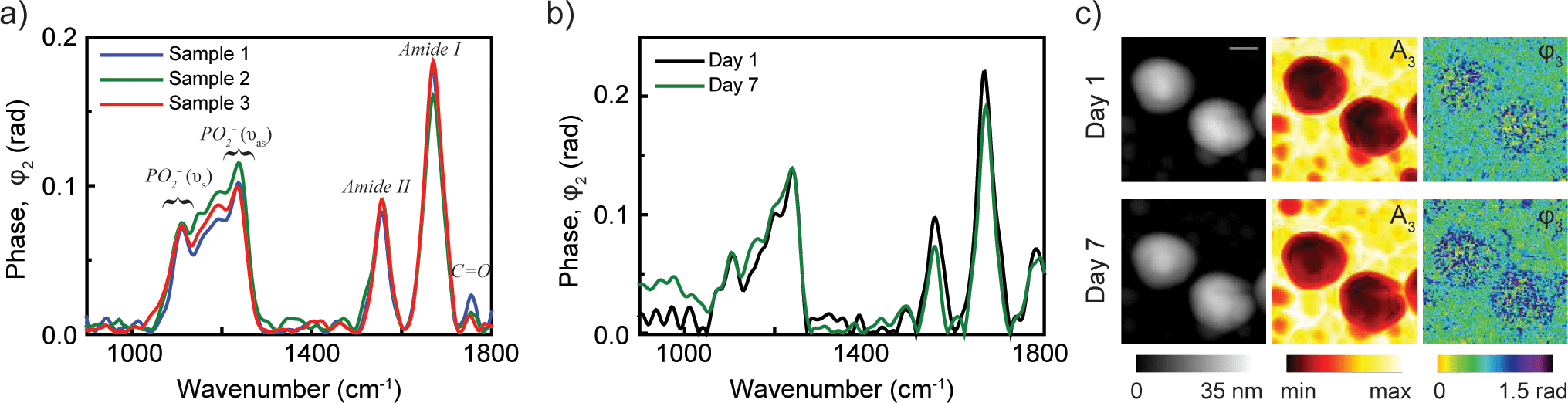
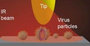
Infrared spectroscopy can detect changes in the chemical and structural composition of viral and host cell membranes caused by the process of viral infection at the molecular level. However, the characteristic size of viruses, lipid envelopes, and surface glycoproteins mediating the fusion process is much smaller than the diffraction limit for infrared light, which prevents the study of the behavior of individual “players” of infection. Therefore, it is important to find a tool that can provide spatial resolution at the nanoscale while analyzing mechanical and chemical properties. Researchers Yohannes Abate and Ming Luoused spectroscopic infrared nano-imaging provided by the neaSNOM microscope to study the chemical and structural changes that occur prior to membrane fusion in a single archetypal influenza virus X31 at different pH environments. Thus, it was also possible to quantify the efficacy of an antiviral compound (Compound 136) in preventing viral membrane disruption: a new mechanism for inhibiting virus entry into the cell. Neaspec’s IR nanoimaging and nano-FTIR spectroscopy microscopes provide a unique tool for analyzing the mechanisms underlying the functionality of viral and cell membranes at the nanoscale, which can significantly support advances in basic virus research and treatment development.
")

