Why combine Raman and electron microscopy (SEM)? Electron microscopy can obtain images of your samples with very high spatial resolution and the use of EDS detector and a detailed elemental map of your sample.
Adding Raman Imaging Microscopy will take your results to the next level:
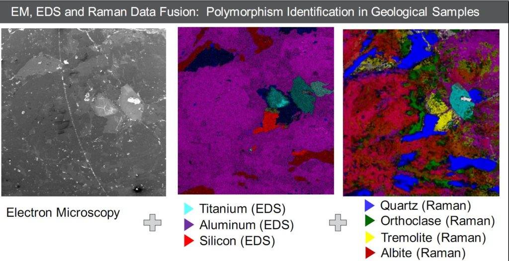
- Samples can be analyzed in parallel with SEM / EDS by Raman microscopy to reveal molecular details from 2D and 3D structures. Cover the chemical image of your SEM structures up to coencoded_tag_open1 microns with single-point measurement or < fast imaging of large areas with the patented Thermo Scientific MAPS Correlative Microscopy Software
- Characterize the chemical composition of your samples, including morphology and polymorphism
- Raman polarization measurements can reveal crystal orientation, making Raman spectral analysis perfect for understanding the structure and functions of materials
- The Raman spectrum can quickly reveal uniformity, purity, thickness, and other symmetric bonding properties in 2D carbon materials such as graphene and carbon nanotubes
- Raman spectral data can also be used to speculate carbon-nitrogen-oxygen bonds to distinguish molecular structures providing additional information to SEM and EDS data.
- Samples can be measured in their native form, eliminating the need for sample preparation. Raman microscopy is confocal, so that depth profiles of multilayer or embedded samples can be obtained without damaging the sample